Interactive Resources for Schools


This topic takes on average 55 minutes to read.
There are a number of interactive features in this resource:
 Biology
The retina of the eye contains many light sensitive cells. They all contain chemicals which change when light falls on them, triggering an impulse in the sensory neurones which make up the optic nerve. The impulses travel along the optic nerve to the visual areas of your brain. The sensory cells then use energy produced by aerobic respiration in the mitochondria to return the chemicals to their original state.
There are two main types of sensory cells on the retina:
The rays of light coming into the eye from a close object for example a book you are reading tend to be travelling differently to the rays of light from an object away in the distance.
The rays of light which reach your eye from a close object tend to be spreading out – they are diverging. As a result, they need a lot of bending to bring them into focus on your retina.
The rays of light which reach your eye from a distant object are travelling in almost parallel rays. As a result they need less bending to bring them into focus on your retina.
All of the light which comes into your eye passes through the cornea and aqueous humour which bend it towards the retina. However it is the lens which makes sure you can see close up and distant objects equally well. This fine focusing is possible because the lens changes shape.
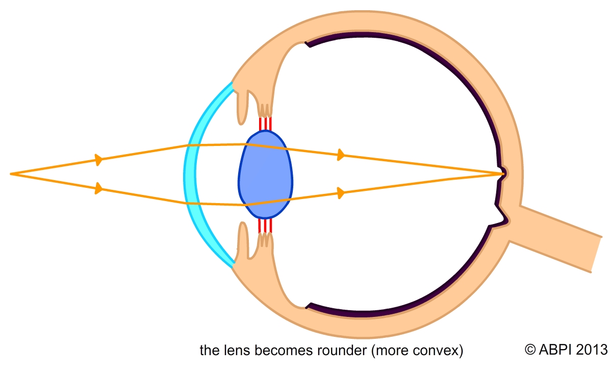
The lens becomes rounder (more convex)
Focusing on a nearby object
Light rays diverging so still need lots of bending when they reach the lens. Lens becomes shorter, fatter and much more convex (rounded) so it bends the light to bring it into focus on the retina.
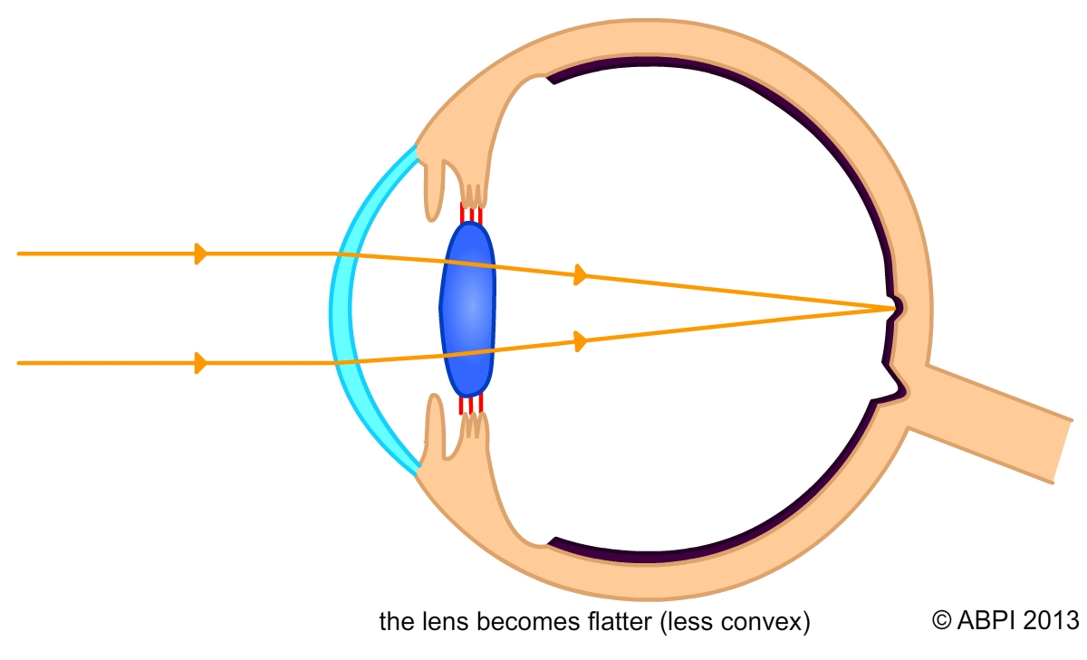
The lens becomes flatter (less convex)
Focusing on a distant object
Light rays almost parallel so need little more bending to bring them into focus on the retina once they have passed through the front of the eye. Lens stretched long, thin and relatively flat so it has little effect.
How does the lens change shape? It is surrounded by circular muscles called the ciliary muscles. These muscles are attached to the lens by the suspensory ligaments. These ligaments do not stretch or give. The ciliary muscles contract or relax in response to the type of light entering the eye:
Accommodation is the term used for the ability of the human eye to focus on objects at different distances away.