Interactive Resources for Schools


This topic takes on average 55 minutes to read.
There are a number of interactive features in this resource:
 Human biology
 Biology
 Physical education
Biology
Physical education
Biology
 Science (applied)
Biology
Science (applied)
Biology
Cells were first discovered in the 17th century as microscopes were developed. Since then, they have been recognised as the fundamental units of the structure, function and organisation of all living organisms. Much of our knowledge and understanding of cells springs from observations of cell structure and ultrastructure using different types of microscopes. Developing microscope technology has played an important role in providing evidence to support hypotheses regarding the roles of cells and their organelles in living organisms.
Light or optical microscopes were developed in the 17th century and are still widely used globally. Light is directed through a sample of biological material and passes through an objective lens and an eyepiece lens to produce an inverted, greatly magnified image focused at the eye.
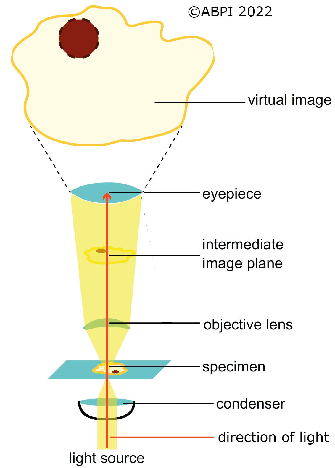
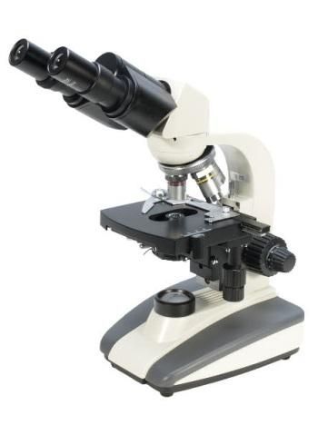
The basic principles of a light microscope (Image on right courtesy of Brunel Mircoroscopes Ltd.)
Microscopes magnify specimens making them appear bigger so we can see details that are invisible to the naked eye. There are two factors which affect what we can see through any particular microscope:
Magnification - how much bigger the image we see is than the actual object. This can be calculated in two different ways, depending on the data we have:
(1) Magnification = image size / object size
For example: image size = 100 mm, object size = 0.05 mm →
Magnification = 100 mm / 0.05 mm = x2,000
The magnification you use is a multiple of the magnification of the eye piece lens and the objective lens you select and can be calculated like this:
(2) Magnification = eyepiece lens x objective lens
For example: eyepiece lens = x10, objective lens = x10 →
Working magnification = 10 x 10 = x100
In a light microscope, magnification is limited by the wavelength of light and the resolving power of the microscope. When you are working out the magnification, make sure all your measurements have the same units – don’t mix micrometres, nanometres and millimetres.
|
Remember : |
1000 nanometres (nm) = 1 micrometre (µm) 1000 µm = 1 millimetre (mm) 1000 mm = 1 metre (m) |
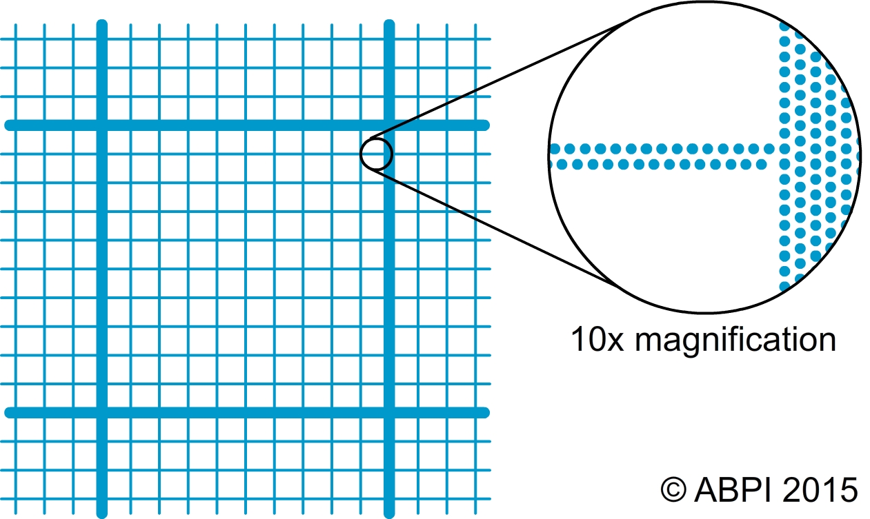
Resolution – the shortest distance between two points that allows the points to be seen as separate items. If they are closer together than this minimum distance, they merge and are seen as a single object. The smaller the limit of resolution, the greater the resolving power of the microscope.
The limit of resolution of the human eye is around 0.1 mm. The print on a page is a mass of tiny dots but you see letters as complete lines because you cannot resolve the dots individually.
To compare the magnification and resolving power of different types of microscopes see the animation at the end of this page.
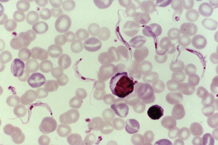
There are many types of light microscopy. They include:
|
A light micrograph of a rat blood smear containing Trypanosoma lewisi parasites (CDC/Dr. Mae Melvin) |
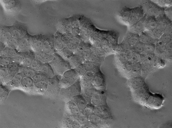
A light micrograph of cells of the rat hepatoma cell line H4IIE at 400x magnification (Shinryuu/CC01.0) |
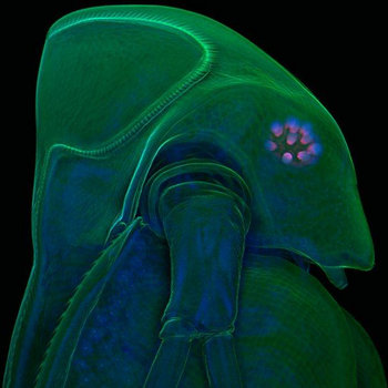
A laser scanning confocal micrograph of a water flea Daphnia atkinsoni (Jan Michels/CC BY-NC-ND 3.0) |
We use different types of light microscopes to provide us with different information about biological material.
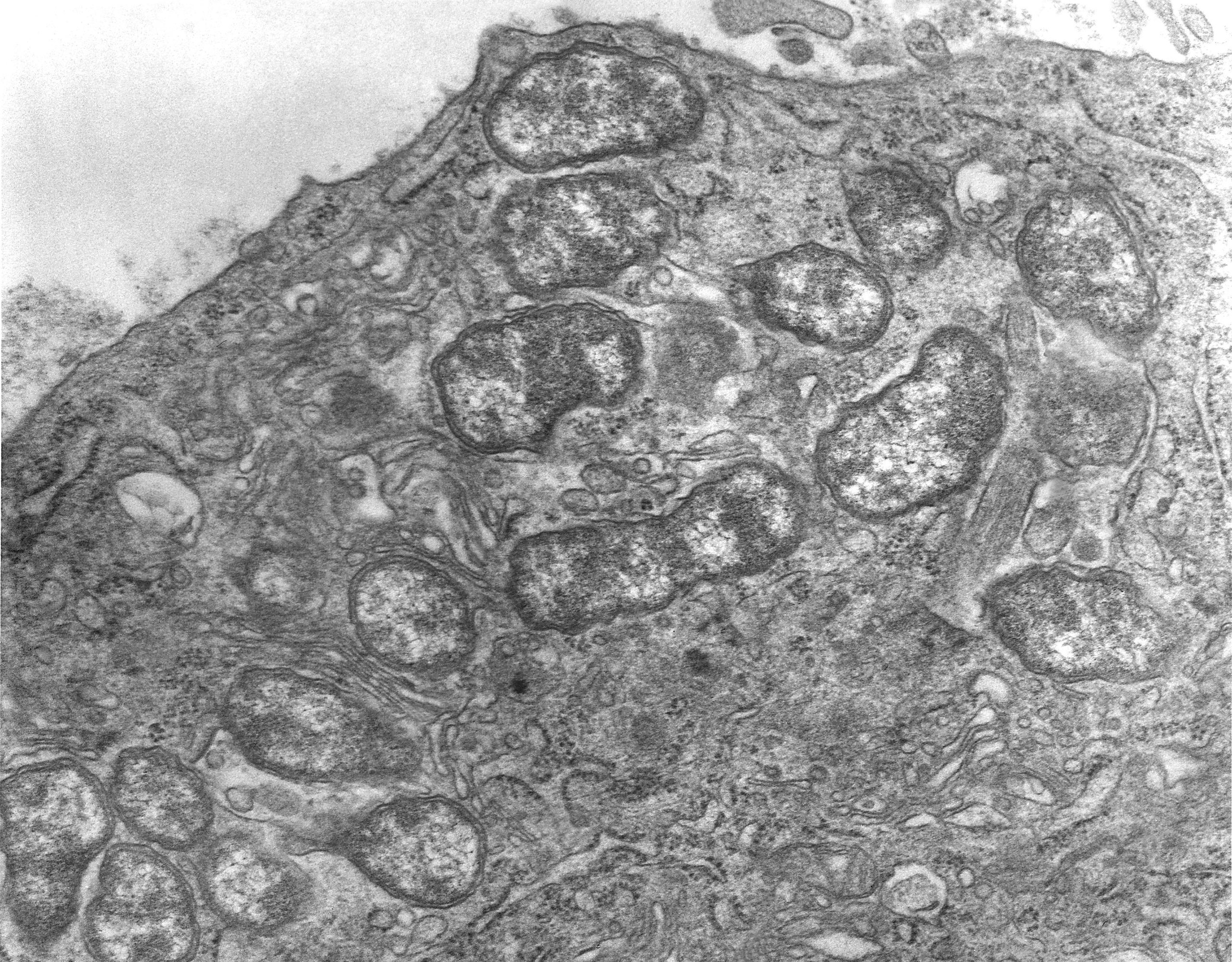
Electron microscopes were developed in the mid-20th century and are widely used in universities and medicine and they have revolutionised our understanding of cells. The image in an electron microscope is formed as electrons are scattered by specially prepared biological material. The electrons behave rather like light waves but with a much smaller wavelength, giving much greater magnification and resolution of the images. The images are always formed on a screen or computer monitor.
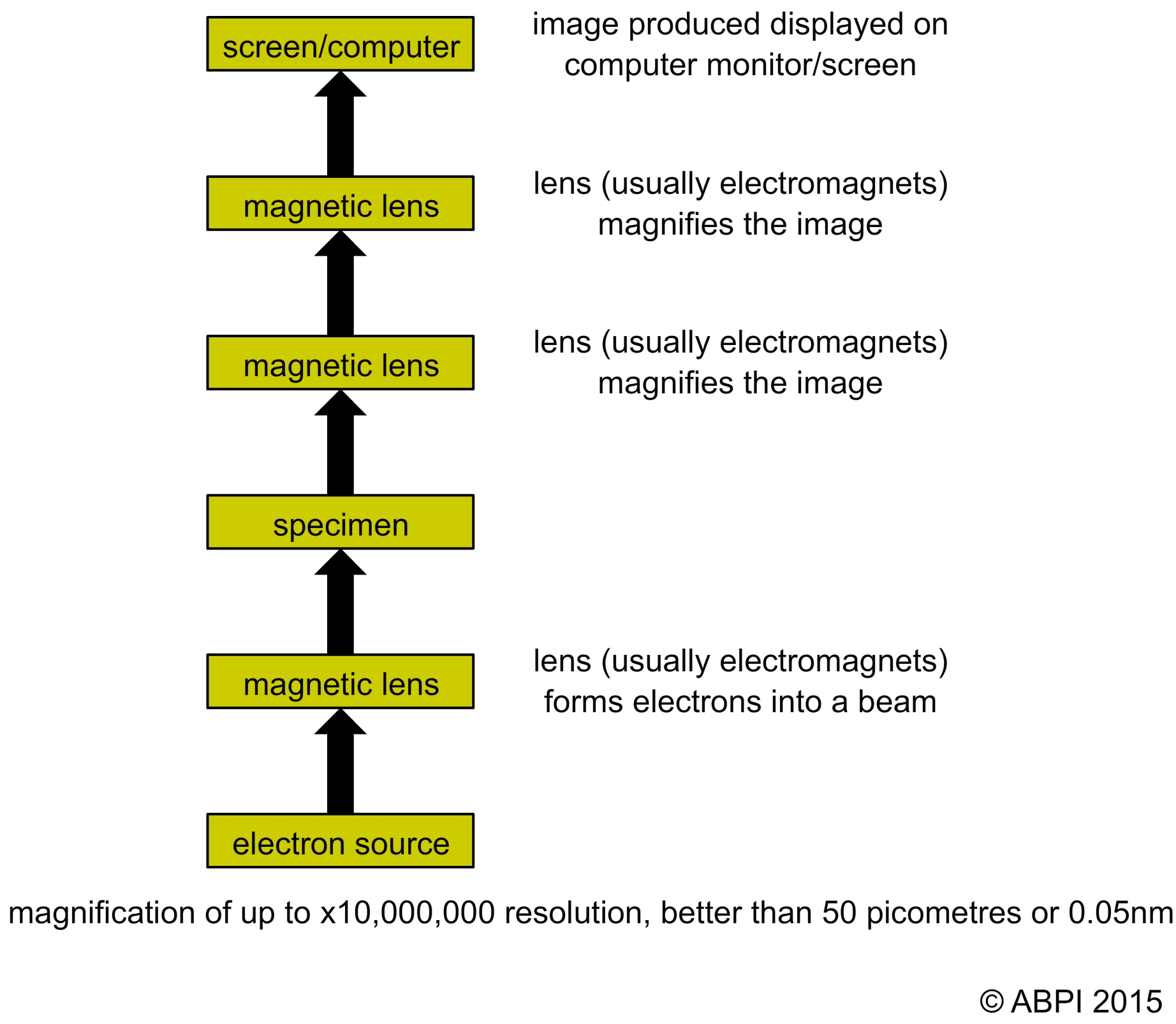
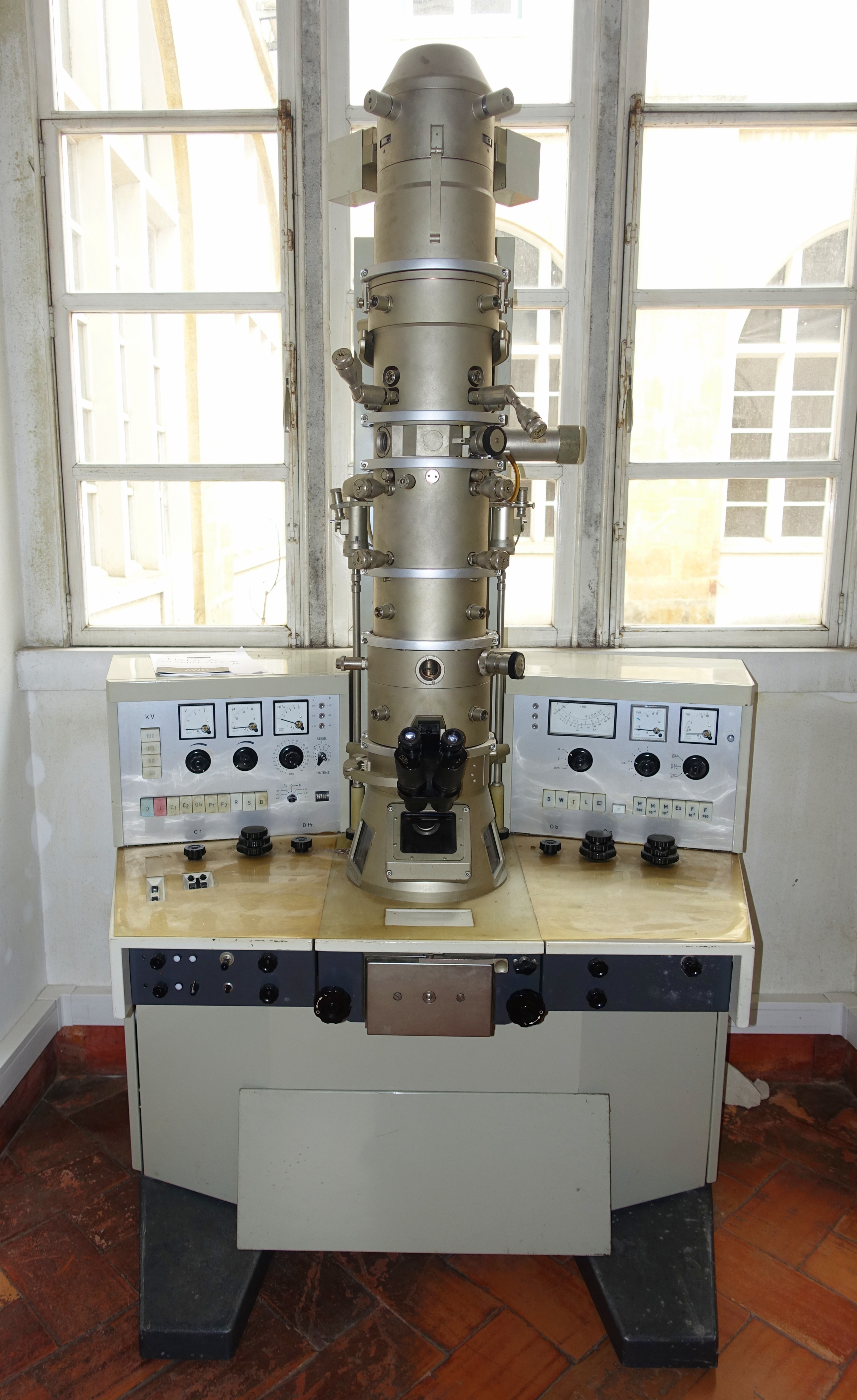
The basic principles of an electron microscope shown next to an electron microscope.
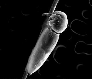
The transmission electron microscope (TEM) gives the highest magnification and resolution and it is used to observe the internal ultrastructure of cells. The scanning electron microscope is used to visualise the surfaces of cells and even whole organisms. The magnification and resolution are lower but the images are three dimensional (3D) and can be spectacular. Transmission and scanning electron micrographs give us very different information about organisms.
To learn more about microscopes, click on the different sections below: