Interactive Resources for Schools


This topic takes on average 55 minutes to read.
There are a number of interactive features in this resource:
 History
 Biology
X-rays were discovered in 1895 by the German Physicist Wilhelm Roentgen. He was studying cathode rays produced by a recently-invented piece of equipment called a Crooke's tube when he noticed that a fluorescent screen across the room started to glow.
The glow appeared to be caused by some unknown rays coming from the Crooke's tube. He tried to block these rays with cardboard but found that they passed straight through it. Amazingly, if he put his hand between the tube and the screen, he could see an image of the bones in his hand. He named this new radiation X-rays and immediately realised how important his discovery would be to the world of medicine.
We are now familiar with atoms, protons, neutrons and electrons but when Roentgen discovered his mysterious rays the structure of the atom was unknown. For this fantastic discovery, Roentgen received the first Nobel prize in Physics in 1901.Today, the use of X-rays is common in hospitals and dentists surgeries all over the world.
We now understand X-rays to be part of the electromagnetic spectrum with a very high energy and short wavelength.
Their high energy means that X-rays can pass through skin and muscle. They are absorbed by dense tissues like cartilage and bone. This produces an image on photographic film which can show damage hidden to the naked eye.
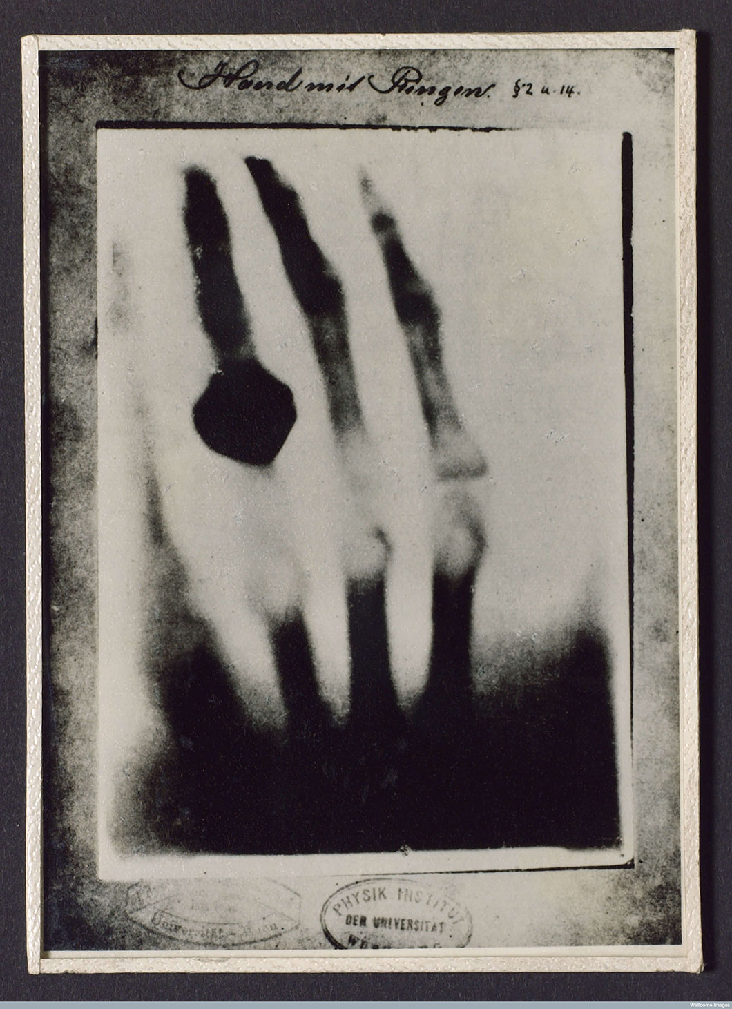
An X-radiograph produced in 1895 by Roentgen of his wife's hand
|
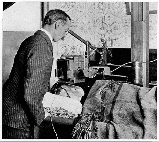
X-ray machine in 1902 |
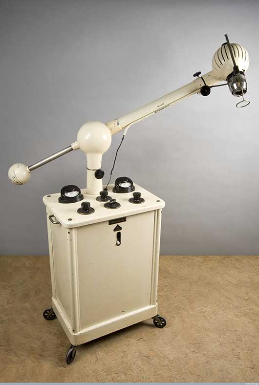
X-ray machine from the 1930s |
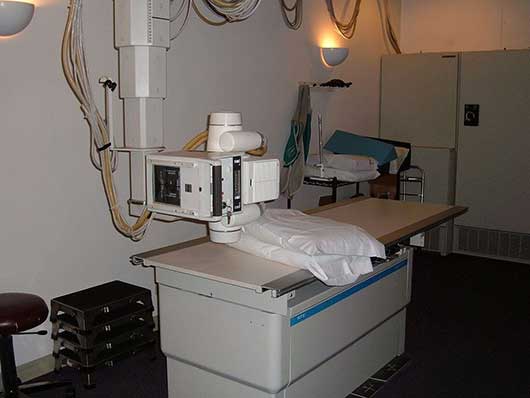
Modern X-ray machine |
A modern development of the X-ray is found in CT scanners. Computerised tomography (or CT) scanners use several beams of X-rays simultaneously from different angles. Detectors measure the amount of each beam that is absorbed in the body and the data is fed into a computer that can build up a virtual, three-dimensional model of the area being examined. CT scans are very sophisticated and can be used to look for damage in much more than bone. They are often used to diagnose and monitor conditions and locate tumours before radiotherapy.
Other forms of imaging in constant use today are Magnetic Resonance Imaging (MRI) and Positron Emission Tomography (PET).
All types of body scanning support accurate diagnoses which help with the choice of different drugs to treat diseases.
X-rays can be used to treat disorders like cancers. In this case, a radiotherapist carefully calculates the dose of X-rays targeted onto the tumour inside the body. These X-rays are over 150 times stronger than those used to in X-radiographs and kill the cancer cells in the tumour.
Low energy X-rays are now widely used to see inside baggage in airports and other areas where security is important.